Watch this to learn about Exercise Cardiac Stress Test with Imaging. You will get information about why a doctor might request one, and what information they can gain from it. It gives your healthcare provider more information than a non-imaging stress test.
This is a way to learn more about your heart. With this type of monitoring, you'll wear a device that records your heart's activity for a period of time as you do different things. Your doctor uses this data to see if there's a problem with your heart.
This test maps blood flow through the heart while you are at rest and while you are doing physical activity. It can show problems with the structure and function of your heart. And, it helps diagnose heart disease or other problems.
This test uses sound waves to make images of your beating heart. We do this test to look for problems with your heart and circulatory system.
This test, which we call an "echo," makes images of your heart. It shows more detail than we see with an x-ray. It lets your doctor see how your heart beats. It can reveal problems within your heart.
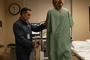
A transthoracic echocardiogram (echo) is an imaging test. It helps your doctor assess your heart. Here's how it works.
An EPS closely monitors your heart rhythm. EPS can help find out exactly what your rhythm problem is and what can be done to control it. A specially trained doctor (electrophysiologist) does the procedure in an EPS lab.
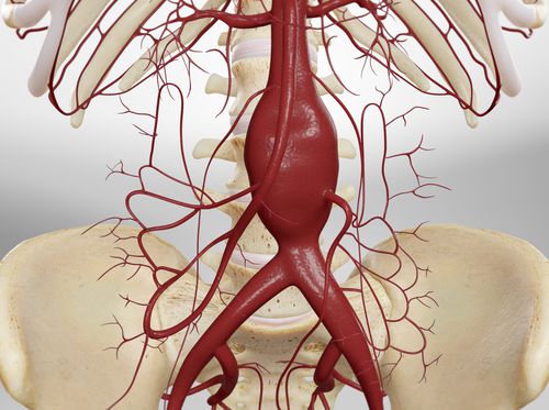
This strengthens a weakened, bulging aorta. That's a large artery in your abdomen. You'll have one or more flexible tubes, called "stent grafts," put in your aorta to support its walls.
This procedure treats a condition called "stenosis." That's when a valve in your heart is narrower than it should be. The valve's flaps, called "leaflets," may be stiff or fused. Balloon valvuloplasty widens the valve so blood can flow through your heart normally.
In this minimally-invasive procedure, a catheter equipped with a balloon and cutting device is used to remove plaque from an obstructed coronary artery. The coronary arteries are the arteries that provide blood to the heart muscles. Directional atherectomy is typically most appropriate for the removal of softer types of plaque.
This is a way we remove plaque that's blocking a coronary artery. The coronary arteries are tiny blood vessels that provide blood to your heart tissue. Clearing a blockage lets your heart get the blood it needs to work properly.
This is a way we remove hardened plaque that's blocking a coronary artery. The coronary arteries are tiny blood vessels that provide blood to your heart tissue. Clearing a blockage lets your heart get the blood it needs to work properly.
In this minimally-invasive procedure, a catheter equipped with a rotating extraction device is used to remove plaque from an obstructed coronary artery. The coronary arteries are the arteries that provide blood to the heart muscles.
This minimally-invasive procedure is used to treat a cerebral arteriovenous malformation (AVM), a tangle of enlarged vessels that can potentially hemorrhage and cause a devastating stroke. In this procedure, fast-drying glue or a small, soft metal coil is placed inside the AVM to help block the flow of blood and prevent hemorrhage.
Watch this video to learn what catheter ablation for atrial fibrillation is and how to prepare for the procedure.
Catheter ablation can fix a cardiac arrhythmia when medicines used to treat it don't work. The ablation stops abnormal electrical signals in your heart. This lets your normal heart rhythm take over again.
This procedure corrects an abnormal heart rhythm. It uses energy pulses to change the way your heart's electrical signals flow.
This test sends a long, thin tube called a "catheter" into the right side of your heart. Through this tube, we get a lot of info about how well your heart is working.
TAVR is a procedure to replace a diseased aortic valve using minimally invasive methods.
Watch to learn the facts behind some common concerns you may have about coronary artery procedures.
Watch how this procedure shows your doctor where and how much your arteries are narrowed or blocked.
This is a procedure used to examine your coronary arteries. Those are the vessels that supply blood to the heart muscle. This procedure can help your doctor find blockages in these arteries.
Learn what you need to do to prepare for an angiography, and what occurs during the procedure.
Learn the possible risks associated with this procedure.
Watch how arteries in your heart become too narrowed and blocked, and how this procedure can restore blood flow.
This procedure uses a balloon-tipped catheter to open one or more arteries in your heart. After an artery is opened, a mesh stent may be placed to hold it open.
Learn how to prepare yourself for your angioplasty, and watch what occurs during the angioplasty and stenting procedures.
See how two types of this medical device are used to improve the health of your heart.
This procedure puts a mesh stent into a narrowed or blocked artery in your heart. The stent widens the artery and holds it open. This improves blood circulation to your heart's tissue.
Learn the possible risks of angioplasty and stenting.
Become familiar with the medications you may receive and the restrictions you will have during your brief stay in the hospital.
Understand how you can avoid complications and safely recover from angioplasty.
Learn the importance of knowing the medications you have been prescribed and how to take them safely.
Learn what signs and symptoms are normal during your recovery, and which ones require immediate attention.
Watch how this possible complication of angioplasty develops, and how to recognize its signs and symptoms.
Angiography is a special type of x-ray that allows your coronary arteries to be viewed and recorded on film. Your doctor can see if the blood vessels to your heart are clogged.
Angioplasty relieves symptoms of coronary artery disease by improving blood flow to your heart. Read on to learn what to expect before, during, and after the procedure.
This nonsurgical procedure widens a narrowed aorta. During the procedure, a balloon is inflated inside the narrowed aorta to expand the blood vessel. Widening the aorta will improve blood flow to the body.
A stent is a small metal coil or mesh tube that is placed in a narrowed artery through a catheter (a long, thin tube) to help improve blood flow to your heart. The stent permanently holds the passageway open and helps reduce the rate of restenosis, renarrowing of the artery.
Watch this to learn what a pacemaker is.
The body generates electrical impulses that cause the heart to beat. In some people, those electrical impulses don't happen in a normal pattern, which can cause the heart to beat too slowly, too fast or irregularly. A very slow heartbeat can lead to fatigue, lightheadedness, dizziness and fainting. In patients who are at risk for a very slow heart rate, doctors often recommend a pacemaker.
Watch this video to learn the different types of pacemakers and how to prepare before the procedure.
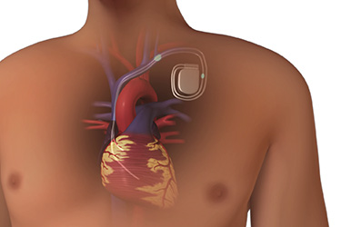
A pacemaker is a small device implanted in the chest to regulate the beat of the heart. Pacemakers may be used to treat a variety of heart problems, including irregularities of the heart's rhythm (called arrhythmias) and weakness of the heart muscle.
Disorders in the heart's electrical system can lead to arrhythmias, or an abnormal heart rhythm. Some patients who have been diagnosed with a slow heart rate, known as bradycardia, may require a pacemaker. A pacemaker is a device that is implanted in the chest and can correct a slow heartbeat.
The procedure for implanting a pacemaker is often routine and relatively easy for the patient. The health care team will tell the patient how to prepare for surgery and what to expect during the procedure.
In this procedure, an impulse generator (called a pacemaker) is implanted in the chest to regulate the rhythm of the heart.
Following the pacemaker implant procedure, the patient will stay in the hospital for one or two nights to make sure the wound is healing without complications and the device is working properly. Before discharging the patient, the health care team will provide instructions on how to care for the wound at home.
Most patients with a pacemaker can live a normal, active life. Patients do need to take some precautions such as carrying their medical ID card, which contains information about their device, and telling all of their heath care providers that they have a pacemaker.
If you've recently been given a pacemaker, you may have some concerns. You may worry that it will keep you from activities you enjoy. But it won't. Sure, you'll need to cut back on some things while your pacemaker settles into place. But soon, you'll be as active as anyone else your age.
Watch this clip to understand how a pacemaker has helped Patsy after her heart attack.
If your heart beats too fast or doesn't beat with a regular pattern, you may need electrical cardioversion. During this hospital procedure, your heart is shocked with electricity. It can help give your heart a normal beat. Cardioversion isn't the same as defibrillation. That's an emergency procedure that uses high-energy shocks. Cardioversion uses low-energy shocks.
A pacemaker is a small electronic device that helps your heart's electrical system beat at the right pace. Inserting the pacemaker into your body is called implantation. You stay awake during the procedure.
You can usually do almost everything you did before you got your pacemaker. See your doctor regularly to help ensure that you remain healthy and feeling good. Here are some things to avoid.
Watch this video to learn what an IDC or implantable cardioverter-defibrillator is and how it's good for your heart.
Watch this to learn what an ICD is.
The body generates electrical impulses that cause the heart to beat. In some people, those electrical impulses don't happen in a normal pattern, which can cause the heart to beat too slowly, too fast or irregularly. A very fast heartbeat can lead to ventricular tachycardia, a potentially life-threatening condition. In patients who are at risk for ventricular tachycardia, doctors often recommend an implantable cardioverter defibrillator or ICD.
Disorders in the heart's electrical system can lead to arrhythmias, or an abnormal heart rhythm. Some arrhythmias can be life threatening and require an electrical shock to return the heart to a normal rhythm. Patients who are at risk for sudden cardiac death may require an implantable cardiac defibrillator or ICD, which can deliver a life-saving shock if the heart starts to beat too fast or in a chaotic pattern.
With this procedure, a small device is placed in your chest. It monitors your heart's rhythm. If it detects that your heart isn't beating normally, it sends electricity to your heart to convert your heart rhythm to a normal one.
This device, which we call an "ICD," is put inside your chest or abdomen. It's used to treat arrhythmia. That's an irregular heart rhythm. An ICD monitors your heart and keeps it beating properly.
Watch this to learn answers to common concerns about getting a heart rhythm device.
The procedure for implanting an ICD is often routine and relatively easy for the patient. The health care team will tell the patient how to prepare for surgery and what to expect during the procedure.
Following the ICD implant procedure, the patient will stay in the hospital for one or two nights to make sure the wound is healing without complications and the device is working properly. Before discharging the patient, the healthcare team will provide instructions on how to care for the wound at home.
Most patients with an ICD can live a normal, active life. Patients do need to be aware of the types of shocks an ICD delivers and what to do if they experience a shock. There are also some precautions patients should take in order to reduce complications.
An ICD is a device that is placed permanently inside your body. An ICD monitors your heart rhythm (the speed and pattern of your heartbeat). If this rhythm becomes too fast or too slow, the ICD sends out electrical signals that help bring the rhythm back to normal. Read on to learn more.
Watch how the angioplasty procedure restores blood flow in the narrowed peripheral artery.
Watch this checklist to help prepare for your angioplasty.
Watch this checklist to help understand what you can do to help your recovery from angioplasty at home.
This is a treatment for peripheral artery disease in the legs. It improves blood flow through an artery clogged with plaque.
Watch how femoral bypass surgery restores blood flow in a blocked artery.
Watch what you will need to do to help prepare for femoral artery bypass surgery.
See how you can help recover at home from your femoral artery bypass surgery.
Watch how this procedure clears blockages and restores blood flow in the arteries of your arms and legs.
Watch what you will need to do to prepare for your atherectomy.
See what to expect as you recover from the atherectomy procedure.
This procedure removes plaque that's blocking a carotid artery. That's an artery that sends blood to your brain and your face. You have two of these arteries, one on each side of your neck.
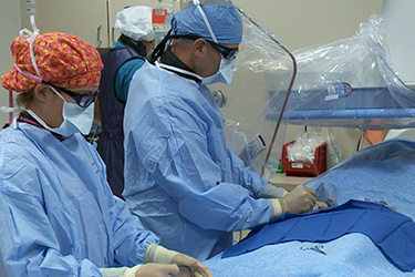
This surgery replaces a defective valve in your heart with one that works properly. The new valve may be mechanical. Or, it may be a valve taken from a human or animal donor.
This is a procedure to fix a problem with your heart's mitral valve. That's the valve between the two chambers on the left side of your heart.
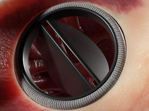
Heart valves regulate the flow of blood through the heart. If a poorly-functioning valve cannot be repaired, it may be replaced with a mechanical or biological valve. Any of the four heart valves can be damaged, but the mitral and aortic valves are the ones most frequently replaced. This animation will show the replacement of the mitral valve through a small opening in the patient's side.
This procedure treats aortic valve stenosis. It replaces your damaged aortic valve with a new valve. We'll use a tube called a "catheter" to put in the new valve. That's easier on your body than using surgery to open your chest and heart.
Learn what to expect before, during, and after heart valve surgery.
For the first 6 to 8 weeks after heart valve surgery, you'll gain a little more energy and strength each day. Your healthcare provider will discuss what you can and can't do as you recover. Here's what you can expect.
Valve disease occurs when a valve doesn't open or close the way it should. If a valve doesn't open all the way, the heart has to push blood through a smaller opening. If the valve doesn't close tightly, some blood will leak backward.
Aortic stenosis means your aortic valve has a problem opening. The left ventricle has to work harder to push the blood through the valve. In some cases, this extra work will make the muscle of the ventricle thicken. This type of stenosis can quickly get worse.